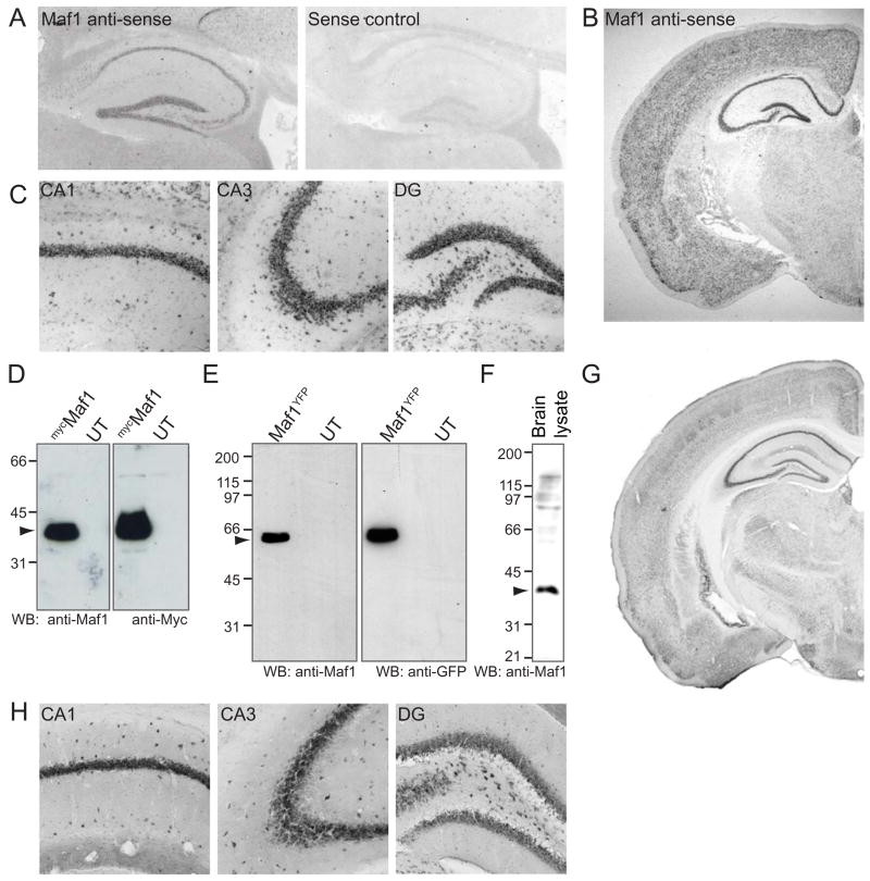
Figure 1. Maf1 is highly expressed in the hippocampus and cortex.
(A) In situ hybridisation of mouse parasagittal cryosections with Maf1 anti-sense or sense control DIG-labelled riboprobes showing Maf1 RNA localisation in the hippocampus. (B,C) In situ hybridisation of whole brain showing Maf1 localisation in the cortex. Close-up images of hippocampus showing Maf1 localisation in CA1, CA3 and dentate gyrus. (D–H) Characterisation of Maf1 antibody. (D) Lysates of COS cells expressing mycMaf1 were probed with anti-Maf1 and anti-Myc antibodies demonstrating the specificity of the Maf1 antibody. (E) Lysates of COS cells expressing Maf1YFP were probed with anti-Maf1 and anti-GFP antibodies demonstrating the specificity of the Maf1 antibody. (F) 50 μg brain lysate probed with anti-Maf1 identifies a band of approximately 35 kDa. (G,H) Immunohistochemistry of mouse brain sections with anti-Maf1, showing a high level of Maf1 expression in the hippocampus, specifically in CA1, CA3 and the dentate gyrus (H).

