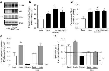
Fig. 2.
Substrate transporters and transport in HL-1 cardiomyocytes. a HL-1 cells were transfected with either pCMV (control) or pCMV-GLUT4myc 24 h after seeding. After a further 48 h cells were lysed and the lysates separated by SDS-PAGE and blotted for GLUT4, myc and CD36. Caveolin 3 (Cav3) served as a loading control. Representative western blots are shown. b, c At 48 h after transfection, serum-depleted cells were treated with 200 nmol/l insulin, electric-field stimulation or 1 μmol/l oligomycin for 30 min. Afterwards, plasmalemmal GLUT4myc (b) and CD36 (c) were detected by immunostaining. d, e Serum-depleted cells were incubated with DMSO (control), 0.2 mmol/l phloretin or 0.5 mmol/l SSO for 30 min. Unbound SSO was washed away and cells were stimulated with 200 nmol/l insulin for 30 min in the continued presence of phloretin (where indicated). Afterwards, the cells were incubated with a substrate mix containing 2-deoxy-d-[3H]glucose and [14C]palmitate for 10 min. (d) Glucose uptake and (e) palmitate uptake were quantified by scintillation counting of the cell lysates. The uptake component that was not inhibited by phloretin, i, presents non-protein-mediated uptake (i.e. passive diffusion), and the uptake component that was not inhibited by SSO, ii, presents the component of palmitate uptake that was independent of CD36. Data are mean values ± SEM of three independent experiments (n=3), with triplicate measurements for each condition. *Statistically different from corresponding basal value (p < 0.05)

