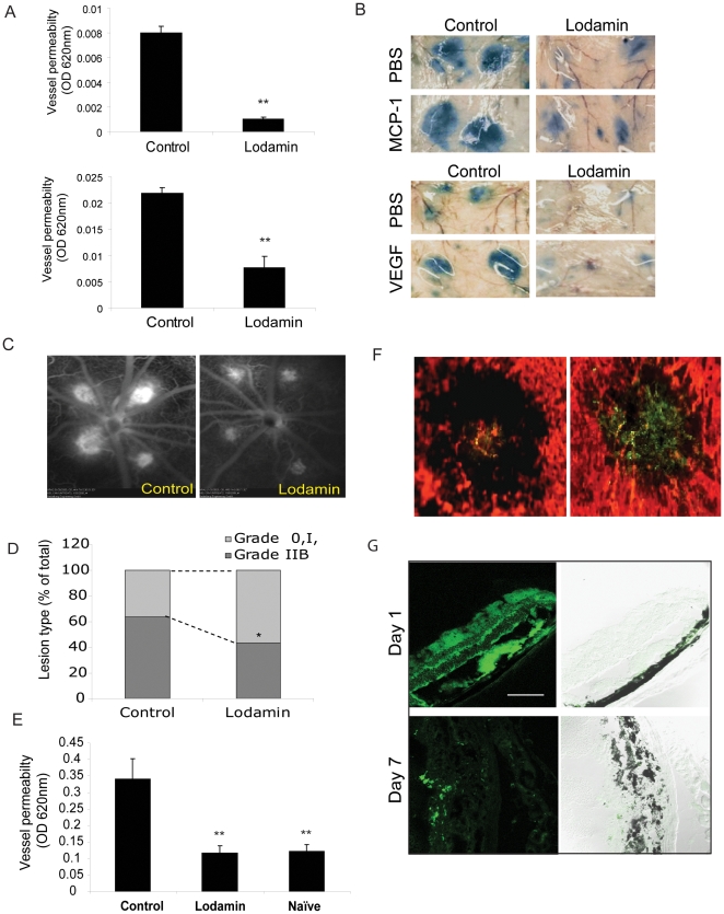
Figure 7. Lodamin reduces vessel permeability in the modified Miles assay and in retinal angiography.
(A) Left: quantification of modified Miles assays performed in mice after induction of vessel permeability using MCP-1 (50 pg) and VEGF (50 ng); extracted dye contents were quantified by measuring at 620 nm. Data are expressed as mean ± SEM (n = 10, *P<0.05, t-test). (B) Representative photos of mouse skin showing diminished dye in mice treated with oral Lodamin 1 and 3 h before conducting the assay. MCP-1 induces vessel permeability in a dose dependent manner, already at 10 pg/ml, MCP-1 induced significant vessel leak compared with PBS. (C) Fluorescein angiography of CNV lesions of rats treated with Lodamin or vehicle. Fluorescein angiography was performed at day 7 after laser photocoagulation and Lodamin was administered 1 and 3 h before performing the imaging. (D) The percentage of lesions graded as 0, I, IIA, defined as no leakage to moderate leakage, and IIB, considered clinically relevant leakage, in vehicle-treated (n = 6 eyes). A significant reduction of vessel leak was observed after an acute treatment of Lodamin. (E) Lodamin reduces whole eye vessel permeability as determined by Evan's blue extraction method. Mouse eyes treated with Lodamin (3 h before conducting the assay) were 65% less than the control. Data are presented as mean ± SD (n = 5, *P<0.05, t-test). (F) Accumulation of 6-coumarin labeled Lodamin injected i.v at the site of CNV lesion in mouse compared to an injection of free 6-coumarin. In red: blood vessels stained with rhodamine concanavalin A, in green: 6-coumarin dye. Bar = 20 µm. (G) Bioditribution of 6-coumarin labeled Lodamin (green) in mouse retina side sections 1 day or 7 days post intravitreal injection. 10 µm Section were imaged by confocal microscopy using optical sections with 488-nm argon, diode lasers. Bar = 20 µm.

