
Figure 6.
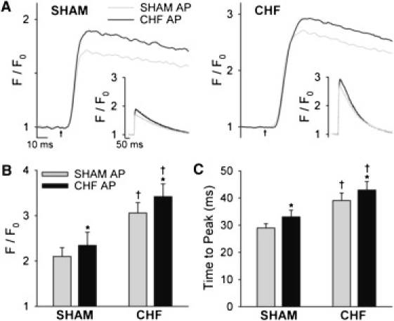
The CHF AP modestly prolonged the peak of the Ca2+ transient. (A) Representative spatially averaged Ca2+ transients from SHAM and CHF myocytes (left and right panels, respectively) during steady-state stimulation with SHAM and CHF APs (insets represent full scale). Mean measurements of Ca2+ transient magnitude (B) and time to peak (C). ncells: SHAM = 9, CHF = 15; ∗P < 0.05 versus SHAM AP; †P < 0.05 versus SHAM cells.