
Figure 1.
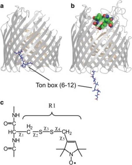
BtuB in the (a) apo form where the Ton box position is highlighted (PDB ID: 1NQE). (b) Vitamin B12 bound form of BtuB showing the state of the Ton box as determined by EPR spectra and pulse EPR distance measurements (based upon PDB ID 1NQH and spectroscopic restraints obtained for the Ton box in bilayers (21)). This unfolding event places the Ton box as much as 30 Å into the periplasmic space. (c) The structure of the spin-labeled R1 side chain and dihedral angles that define the rotamers of R1.