
Figure 2.
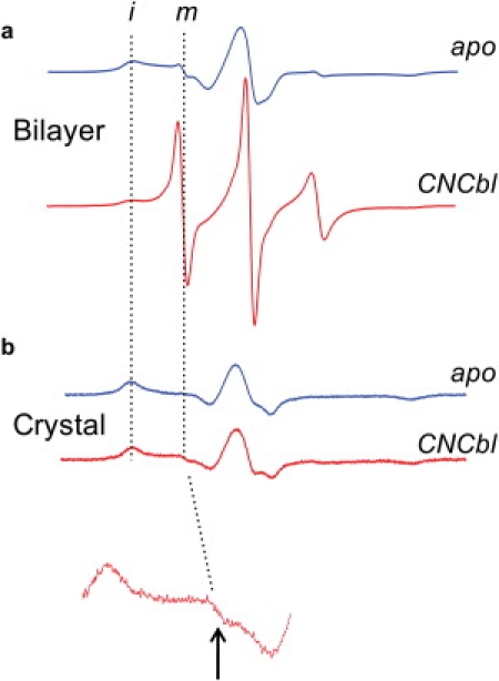
EPR spectra for V10R1 with (red traces) and without (blue traces) substrate when BtuB is incorporated into (a) POPC bilayers, or (b) in the protein crystal. The inset below is a 10× vertical expansion showing a small signal from unfolded Ton box. The dashed vertical lines indicate the positions of signals resulting from immobilized (i) and mobile (m) nitroxide side chain, corresponding to folded and unfolded Ton box, respectively.