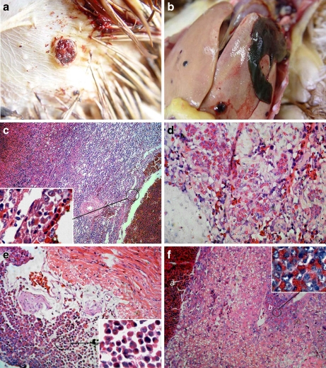
Fig. 1.
Gross lesions and Histopathology (haematoxylin and eosin staining). (a) Hemangioma on the body surface. JS09GY2 case was showed. (b) Hepatorrhexis in JS09GY3 case caused by hemangioma in liver. (c) Spleen of JS09GY6 showing different staged hemangiomas. Myeloid cells aggregations were found around the hemangiomas (H&E stain, 200×). (d) Myeloid cells in the interstitium of the hemangioma of JS09GY2 (H&E stain, 400×). (e) Myeloid cells aggregation in heart of JS09GY5 (H&E stain, 400×). (f) Hemangioma and ML tumor coexisted in the liver of JS09GY3 (H&E stain, 400×), the tumor cells in the ML were observed (inserted, 1,000×); (a) Hemangioma, (b) ML

