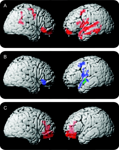
Figure Image analyses of patients with progressive nonfluent aphasia (PNFA) and behavioral-variant frontotemporal dementia (bvFTD)
(A) Significant cortical thinning in PNFA. (B) Regression analyses relating language features to cortical thinning (blue areas indicate regions where reduced WPM is related to PNFA atrophy; green areas indicate regions where the proportion of complex structures is related to atrophy). (C) Significant cortical thinning in bvFTD.

