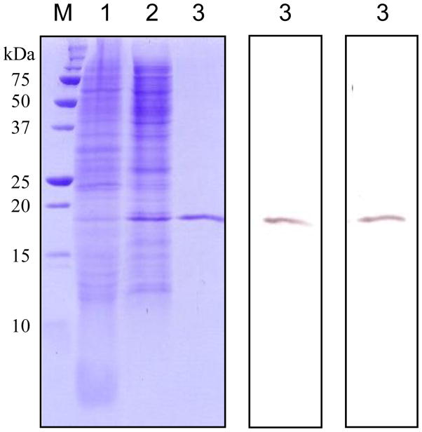
Fig. 3. Isolation of M. sexta PGRP1 from baculovirus-infected Sf9 cells.
Left panel, Coomassie blue staining; central panel, immunoblot analysis using anti-(His)5 as the first antibody; right panel, immunoblot analysis using diluted PGRP1 antiserum as the first antibody. Conditioned cell culture medium (lane 1, 10 μl), proteins eluted from dextran sulfate-Sepharose (lane 2, 10 μl), and affinity-purified PGRP1 from Ni2+-NTA agarose (lane 3, 10 μl) were separated by 15% SDS-PAGE, along with pre-stained molecular weight standards (M) with their sizes indicated on the left.

