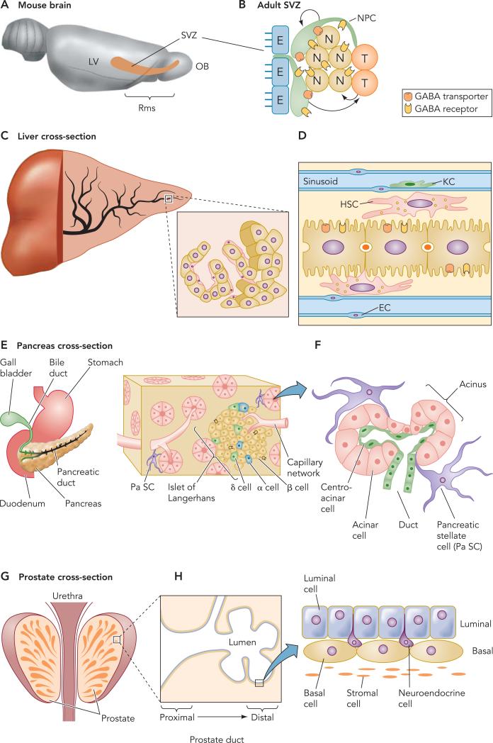
FIGURE 2. The brain and the subventricular zone.
A: sagittal depiction of the brain and the subventricular zone (SVZ)-rostral migratory stream (RMS). B: a diagram of the adult SVZ. Ependymal (E) cells border the lateral ventricle (LV). Astrocytes (Astro) (neural stem/progenitor cells) surround a cluster of neuroblasts (N) and transit amplifying cells (T). Both astrocytes and neurob-lasts have GABAA receptors, and astrocytes also have GABA transporters. C: structure of a portion of a hepatic lobule. D: diagram illustrating the parenchymal (hepatocyte) and nonparenchymal cells in the liver. Endothelial cells (EC) form the lining of the sinusoids (S). Kuffler cells (KC) are tissue macrophages. Stellate cells lie in the space between hepatocytes and endothelial cells. Arrows and asterisks indicate a classical and new definition of the perisinusoidal space of Disse between hepatocyte and stellate cells, respectively, and endothelial cells. GABAA receptors are expressed in hepatocytes. E: diagram illustrating the different cells of the pancreas in the islet of Langerhans (endrocrine pancreas) and the acini (exocrine pancreas). F: diagram illustrating the location of pancreatic stellate cells. G: cross-section of the prostate. H: schematic of basal and luminal cells.

