
Figure 1.
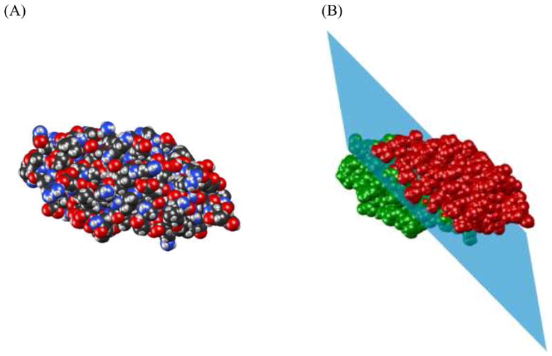
Illustration of the bisection of Cyanovirin-N (PDB code 2EZM). (A) Van der Waals surface of Cyanovirin-N. (B) Illustration of how the protein is split into two domains with approximately equal number of atoms by a plane. The first domain is colored green, the second domain is red.