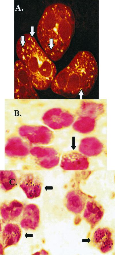
Figure 1.
Determination whether Eos contain organelles that label with mitochondrial-specific dyes and contain mitochondrial-specific DNA. (A) Partitioning of CMX into organelles within Eos. Examination by fluorescence confocal microscopy revealed 24–36 labeled organelles per Eos, as indicated by white arrows. (B and C) In situ PCR amplification and visualization of the mitochondrial-specific gene, cytochrome oxidase subunit II. Peripheral blood cells were used in these experiments. Black arrows indicate Eos. (B) The controls that contained no DNA primers specific for cytochrome oxidase subunit II showed little to no endogenous staining. (C) In situ PCR with DNA primers for cytochrome oxidase subunit II showed 24–36 organelles per Eos. Mitochondria of other cell types were not labeled, because PCR conditions were specifically optimized for labeling Eos. A representative experiment of n = 3 experiments with consistent results.

