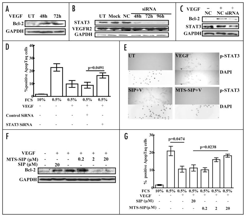
Figure 5.
VEGF induces Bcl-2 and protects endothelial cells from death by a STAT3-dependent mechanism. HUVEC were placed in serum-free medium overnight, stimulated with medium containing 0.5% serum + VEGF (100 ng/ml) for 48 or 72 hours. Blots of the lysates were probed with antibody to Bcl-2, stripped and reprobed with antibody to GAPDH (A). HUVEC were transfected with STAT3 siRNA (siRNA; 100 nM) for 48, 72 or 96 hours, transfected with negative control siRNA (NC; 100 nM) for 96 hours, mock transfected (Mock) or untransfected (UT). Blots were probed sequentially with antibodies to STAT3, VEGFR2 and GAPDH (B). HUVEC were transfected with STAT3 siRNA (siRNA; 100 nM) or negative control siRNA (NC; 100 nM) for 72 hours. The transfected cells were cultured in serum-free medium overnight and then placed in medium containing 0.5% serum + VEGF (100 ng/ml). Cell death was assayed after 24 hours and cell lysates were prepared after 48 hours. Blots were probed with antibodies to Bcl-2 and STAT3, stripped and reprobed with antibody to GAPDH (C). Cell death was assayed by TUNEL staining (D). This experiment was performed a total of three times with similar results. HUVEC were treated with 20 μM p-STAT3 inhibitory peptide linked to a membrane translocating sequence (MTS-SIP) or with unlinked SIP at 20 μM in serum-free medium for 16 hours. They were then stimulated with VEGF (100 ng/ml) for 10 minutes. Cells were fixed and stained using anti-p-STAT3 antibody. DAPI counterstaining of nuclei is shown (E). HUVEC were treated with 0.2, 2 or 20 μM p-STAT3 MTS-SIP or with unlinked SIP at 20 μM in serum-free medium for 16 hours. They were then cultured in medium containing 0.5% serum + VEGF (100 ng/ml) and the peptides. Cell death was assayed after 24 hours and cell lysates were prepared after 48 hours. Blots were probed with antibodies to Bcl-2, stripped and reprobed with antibody to GAPDH (F). Cell death was assayed by TUNEL staining (G). All Western blot experiments were performed twice and the other experiments were performed a total of three times with similar results.

