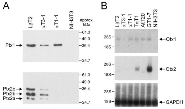
Fig. 6. Expression of Otx and Ptx HD Proteins in Pituitary-Derived Cell Lines.
A, Ptx1 and Ptx2 are expressed in both LβT2 and αT3-1 cells. A, Western blot with approximately 15 μg of nuclear extract from LβT2, αT3-1, αT1-1, and NIH3T3 cells was incubated with antibodies specific to Ptx1 or Ptx2. For Ptx1, a single band is observed in LβT2, αT3-1, and αT1-1 cells. The three bands observed with the Ptx2 antibody represent the three isoforms of Ptx2 (21), as indicated. B, Otx1, but not Otx2, is expressed in gonadotrope-derived cell lines. A Northern blot with approximately 2 μg poly(A) mRNA from LβT2, αT3-1, αT1-1, TαT1, AtT20, GT1-7, and NIH3T3 cells was probed with radiolabeled mouse cDNA fragments coding for Otx1 or Otx2. The GAPDH cDNA was used to allow visualization of the quantity of RNA loaded in each lane. Otx1 is detected in all cell types except NIH3T3, although at low levels in αT3-1 cells. Otx2 is detected in TαT1 and GT1-7 cells but not any of the gonadotrope-derived cell lines.

