
Fig. 3.
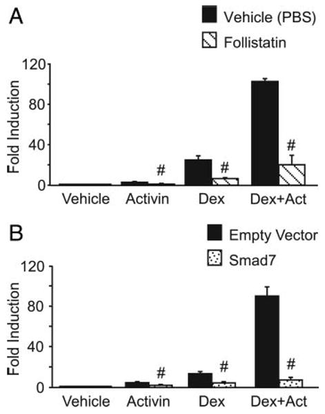
Disruption of activin signaling significantly reduces synergy on FSHβ. The −1000FSHβluc reporter gene and TKβgal were transiently transfected into LβT2 cells along with 200 ng GR. After overnight starvation in serum-free media, the cells were treated for 24 h with 10 ng/ml activin and/or 100 nM dexamethasone (Dex). A, Cells were treated with vehicle (0.1% ethanol), activin, and/or dexamethasone plus vehicle control for follistatin (PBS) and were normalized to the PBS-treated vehicle control (black bars), or cells were treated with vehicle (0.1% ethanol), activin, and/or dexamethasone plus 100 ng/ml follistatin and normalized to the follistatin-treated vehicle control (striped bars). #, Significant reduction with addition of follistatin in comparison with cells without follistatin. B, Cells were treated with vehicle (0.1% ethanol), activin, and/or dexamethasone and were transfected with the empty vector for Smad7 and normalized to the empty vector-transfected vehicle control (black bars), or cells were treated with vehicle (0.1% ethanol), activin, and/or dexamethasone and transfected with 200 ng Smad7 and normalized to the Smad7-transfected vehicle control (dotted bars). #, Significant reduction with overexpression of Smad7 in comparison with the empty vector by Student's t test.