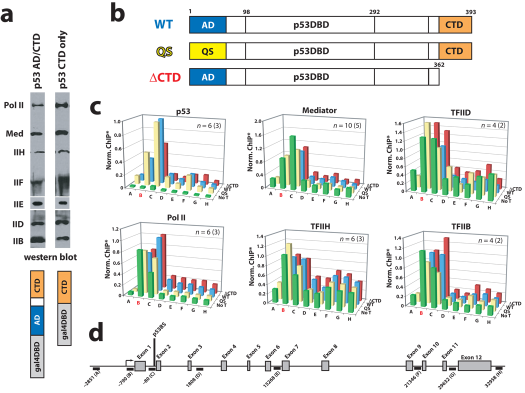
Figure 1.
The p53AD is not required for PEC assembly in vitro or in cells. (a) Immobilized template assays. Occupancy of PEC components in the presence of promoter-bound GAL4–p53CTD or a GAL4–p53AD/CTD fusion protein. Note that endogenous p53 will not bind the GAL4 promoter. Antibodies—pol II: Rpb1; Mediator: Med1; IIH: ERCC3; IIF: Rap74; IIE: IIEα; IID: TBP. (b) Schematic of WT p53 and mutant p53 proteins used in this study. (c) ChIP assays at HDM2 in p53-null HCT116 cells following transfection with wild-type p53 (WT, blue bars), p53 with a truncated CTD (residues 1–362; ΔCTD, red bars), or p53QS mutant (QS, yellow bars). Data from no transfection controls (No T, green bars) are also shown. The probe location in red (B) represents the promoter/transcription start site. Note that PEC factors appear to be pre-loaded at the promoter, as observed previously21. *ChIP output was normalized to WT p53, typically from C primer. For clarity, error bars are not shown but can be viewed in Supplementary Figure 5. (d) Schematic of the HDM2 locus, showing ChIP probe locations.

