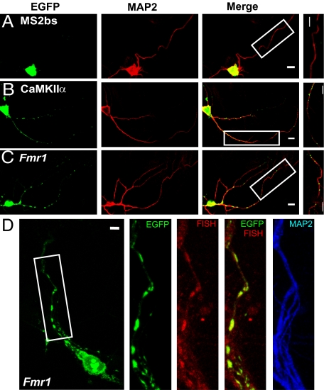
Fig. 1.
Labeling of CaMKIIα and Fmr1 mRNA in primary hippocampal neurons. (A) A neuron transfected with GFP-MS2-nls (nuclear localization signal) and MS2bs showed that GFP signals stay in soma. (B and C) The neuron transfected with GFP-MS2-nls and MS2bs-CaMKIIα or MS2bs-Fmr1 showed that mRNA puncta distribute in dendrites. Higher magnification of the boxed images shows GFP-labeled granules in dendrites. (D) Fmr1-containing GFP-labeled granules (green) in dendrites colocalized with Fmr1 mRNA detected by FISH (red). (Scale bars, 10 μm.)

