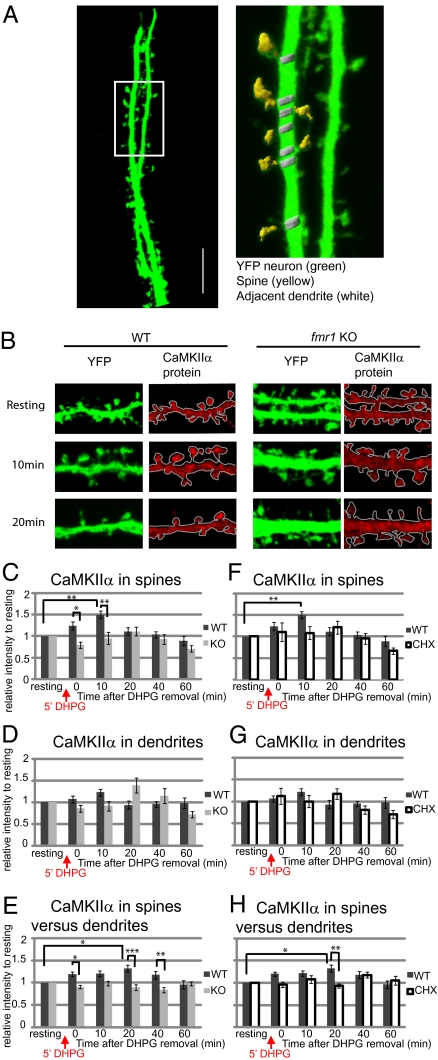
Fig. 4.
The differential distribution of CaMKIIα protein in neuronal spines and dendrites in response to group I mGluR stimulation. Cycloheximide blocks local translation of CaMKIIα in spines of WT neurons. (A) YFP-expressing hippocampal neurons (green) were used to outline neuronal dendrites and spines. A higher magnification of the boxed region shows that spine (yellow) and neighboring dendrite (white) regions could be selected based on YFP staining threshold. (Scale bar, 10 μm.) (B) Representative figures of CaMKIIα immunostaining (red) in WT or fmr1 KO YFP hippocampal neurons show relative CaMKIIα distribution in spines or dendrites in response to group I mGluR stimulation (DHPG). (C and D) The average intensity of CaMKIIα protein in WT or fmr1 KO YFP spines (C) or adjacent dendrites (D) was measured and compared with CaMKIIα level before stimulation. (E) Enrichment of CaMKIIα mRNA in spine relative to adjacent dendrite was calculated and normalized to the level before DHPG treatment. (F and G) In the presence or absence of 60 μM cycloheximide, a protein synthesis blocker, throughout the experiment, the level of CaMKIIα in spines (F) or neighboring dendrites (G) of WT neurons was measured. The relative level of CaMKIIα was standardized to the level before DHPG treatment. (H) In the presence or absence of 60 μM cycloheximide, the level of CaMKIIα in WT spines versus dendrites was calculated as the level of CaMKIIα in one spine divided by the level in the neighboring dendrite. Data were analyzed from at least 18 dendrites in each group from three independent experiments. Statistical analysis by two-way ANOVA with Tukey's HSD posttest. Error bars denote SEM.

