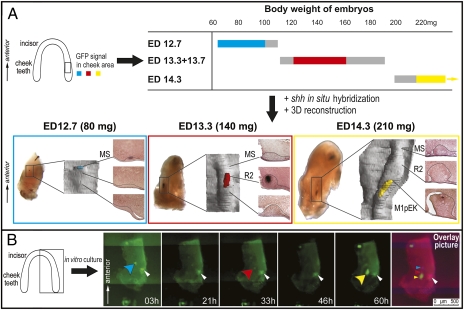
Fig. 2.
Three Shh signaling centers are sequentially patterned in the cheek region of the mandible. (A) The time-table shows in colors (blue, red, yellow) the presence of a distinct signal patch in the cheek region of mandible of the Shh-EGFP mice according to their age (ED) and body weight (mg). Gray represents the mandibles with weak or indistinct signal. Blue, red, and yellow boxes represent the mandibles with the signal patch in the cheek region at ED 12.7, 13.3 + 13.7, and 14.3, respectively. The mandibles hybridized with Shh antisense probe were sectioned frontally to show dental epithelium (in a rectangle), which was reconstructed in 3D (Shh signal in color according to the time-table). The Shh signaling centers correspond to the respective morphological structures MS, R2, and M1 bud. (B) Time-lapse microscopy beginning at ED 12.7 (90 mg), culture time in picture corner. Blue, red, or yellow arrowhead designates three successive EGFP patches; the white arrowhead designates a necrotic zone. An overlay of the pictures at 0, 31, and 60 h shows three distinct EGFP signals.

