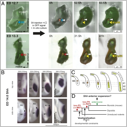
Fig. 4.
Sequential patterning of the cheek teeth in mouse mandible. (A) Time-lapse microscopy pictures of Shh-EGFP mandibles cultured after DiI microinjection at ED 12.7, 90 mg (in MS) or at ED 13.3, 145 mg (in R2), (culture time in picture corner). Note the pEK of the M1 appears posteriorly to the DiI label. The R2 label is later overlapped by the anteriorly extending M1pEK. (B) A series of ED 14.0 dissociated dental epithelia hybridized with Shh antisense probe document the secondary anterior extension of the M1 Shh-signaling at the bud-cap transition. (C) A model of integration of the premolar rudiments in patterning of mouse molars. (Arrows) The influence of a tooth primordium on the newly rising one. (D) A two-step working model of mouse tooth row evolution.

