Figure 5.
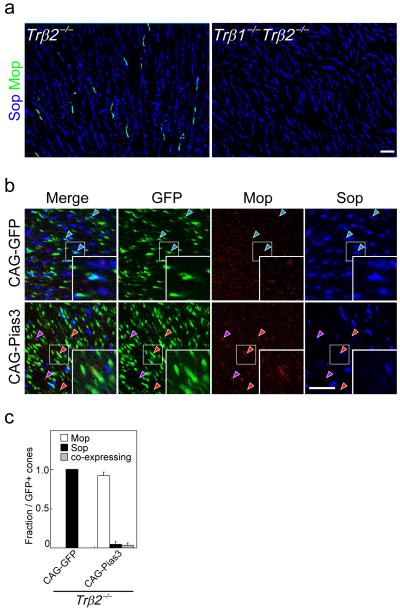
Pias3-dependent regulation of cone subtype specification can occur independently of Trβ2. (a) Flat mount IHC of the dorsal region of 8wk Trβ2−/− and Trβ1−/− Trβ2−/− mouse retinas labeled with antibodies to Sop and Mop. (b) Flat-mount immunohistochemistry of the P14 Trβ2−/− mouse retinas electroporated in vivo at P0 with CAG-GFP and CAG-Pias3 labeled with antibodies to Mop, Sop, and GFP antibodies. Blue arrowheads indicate electroporated S-dominant cones. Red arrowheads indicate electroporated cones that are M-dominant, while purple arrowheads indicate M-dominant cones that do not express detectable levels of GFP. Inset in each panel is a high magnification image of electroporated cones (white box). Purple arrowheads represent non-electroporated cells expressing M-opsin. (c) Composition of electroporated cone subtypes shown in Fig. 5b. All data are represented as mean ± s.d. (n = 3). Scale bar: 20 μm.

