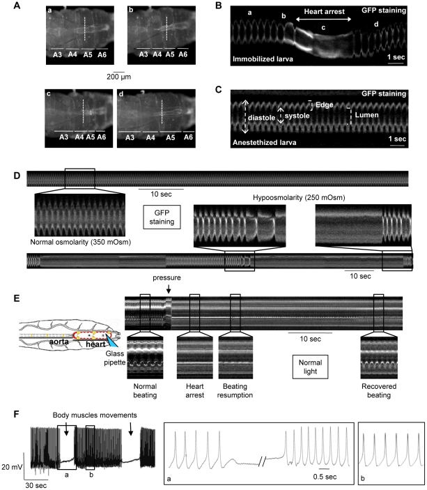
Figure 1. Mechanosensitive response of the cardiac tube in control (y,w; CS ×1029-Gal4) larvae.
Heart movement detection is followed during larval motion of a larva immobilized on double-faced tape. Single movie frames (A, posterior is Right) and m-mode traces (B) are extracted from the Video S3. The m-mode trace, excised at the same position (dotted line) in all movie frames, displays a regular frequency (170 bpm) in absence of larval motion (a). When the larva attempts to crawl, the frequency decreases (b) and a 2 sec-cardiac arrest in diastole occurs during the maximal shortening of the larva (c). The basal frequency is recovered shortly following larval relaxation (d). (C) M-mode trace is extracted from the Video S2 of a typical anaesthetized larva. The cardiac tube displays a regular rhythm (170 bpm). (D) Hypoosmotic shock in semi-dissected cardiac tube. (Top) M-mode trace in normal osmolarity displays a regular rhythm (160 bpm). (Bottom) A 20% decrease in frequency is observed after 10-sec exposition to hypoosmotic solution. One minute after, periodic pauses (30–40 sec) in diastole occur after strong bradycardia and the heart restarts spontaneously at the end of the pause. (E) Direct mechanical pressure exerted on semi-dissected cardiac tube. The cardiac tube is submitted to a local pressure (posterior part of A6 segment) with a fire-polished pipette as indicated in the schematic drawing. The heartbeat is regular before the pressure, then stops when the pressure is applied and restarts slowly 15 sec after the removal of the pipette. Basal frequency is recovered after 30 sec. (F) Action Potential (AP) recording in a semi-dissected cardiac tube. The microelectrode is implanted in the posterior part of the A6 segment. Stable and reproducible APs were recorded with the characteristics previously described for Drosophila larvae [11]. APs stop during body muscles contraction concomitant with cardiac arrest in diastole, and a slow membrane depolarization is recorded until the depolarization phase of the next AP, when the heartbeat restarts after the wave of body muscle contraction. Note that the AP frequency is higher when the activity restarts and returns to normal frequency after 3–5 seconds. In (A–D), cardiac tubes are labeled with NP1029-Gal4-driven membrane bound GFP. In (E), heart movement detection is followed in normal light. In (D–F), preparations are bathed in Schneider medium.

