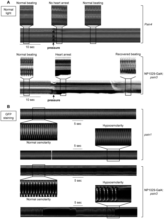
Figure 3. Response to in situ mechanical stress in pain knockdown individuals.
(A) Direct mechanical pressure exerted on semi-dissected pain4 cardiac tube. No modification of cardiac rhythm is observed when the mutant cardiac tube is submitted to a local pressure (applied as described for the wild-type in Figure 1E). This mutant phenotype is rescued by cardiac-specific re-expression of pain in pain3x1029-Gal4 in which the cardiac pause is recovered following the mechanical stimulus. (B) Hypoosmotic shock in semi-dissected cardiac tube. M-mode trace in pain1 mutant displays a regular cardiac frequency in normal osmolarity (210 bpm) preserved after exposure to hypoosmotic solution. This mutant phenotype is rescued by cardiac-specific re-expression of pain in pain3x1029-Gal4 in which the periodic pauses in diastole are observed after strong bradycardia. In (A), heart movement detection is followed in normal light. In (B), cardiac tubes are labeled with NP1029-Gal4-driven membrane bound GFP. Preparations are bathed in Schneider medium.

