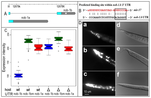
Figure 8. Validation of nob-1 as a target of mir-57.
(A) Two alternative forms of nob-1 3′ UTRs (grey bars) are shown in scale. Blue bars are coding exons and black lines introns. A putative mir-57 binding site within the nob-1b UTR is shown as a white star. (B) Sequence of a putative mir-57 binding site within the nob-1b 3′ UTR (chrIII:12077862-12077884, WS205). The mature mir-57 sequence is highlighted in red. Putative seed sequences are enclosed in the rectangle. (C) Effect of the binding site on the reporter expression. Shown are boxplots of tail expression intensities derived from three different 3′ UTRs: nob-1b, nob-1bm (nob-1b UTR with site-directed removal of the binding site), nob-1a injected into wild type (wt, first three columns) or mir-57 mutant (Δ last three columns) animals as indicated (See Materials and Methods). (D) Micrographs of reporter expression in wild type (a and b) or mir-57 mutant animals (c). Animals were injected with constructs carrying wild type (a) or mutated nob-1b UTR (b). The same array as that used in (a) was crossed into mir-57 mutant animals (c). d, e, f are DIC pictures of a, b and c respectively. Vab tails are caused by mir-57 promoter array.

