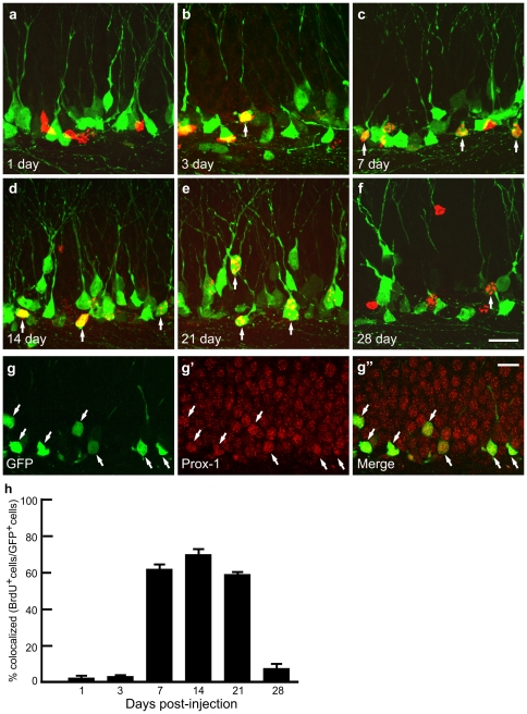
Figure 3. GFP+ granule cells are newborn neurons.
a-f, BrdU-labeled nuclei (red) and GFP expressing cells (green) in the dentate gyrus at various stages after BrdU injection. Arrows show colocalization of BrdU-labeled nuclei and GFP expressing cells. g, All GFP+ cells express Prox-1, a dentate granule cell marker. h, Percentage of BrdU positive cells that are also GFP positive. Percentages of co-labeling are 1.3±0.3% at 1 day, 2.1±0.3% at 3 days, 62.5±3.2% at 1 week, 70.8±3.1% at 2 weeks, 58.7±1.3% at 3 weeks and 7.4±1.9% at 4 weeks (n = 4 mice each time point, mean±SEM). Scale bars, 20 µm.

