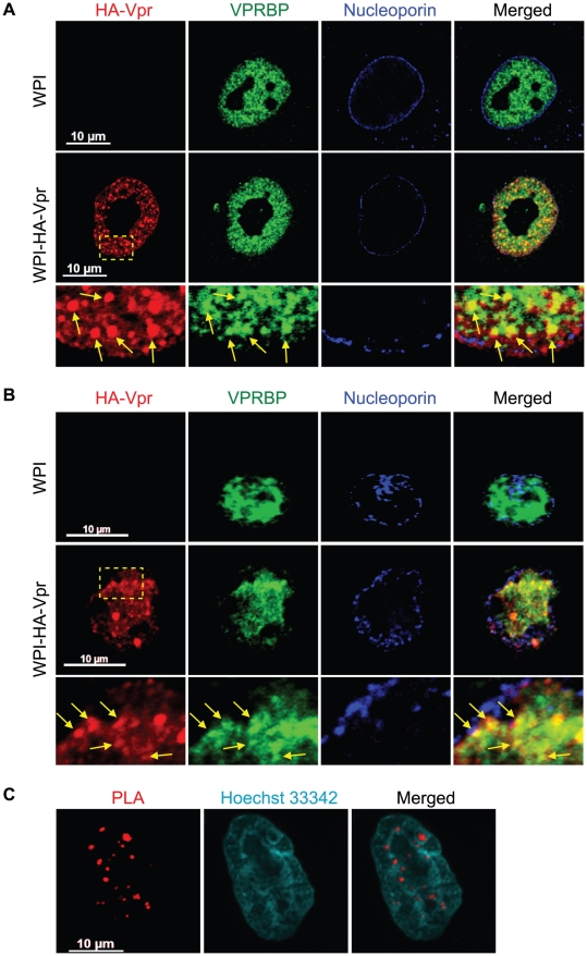
Figure 1. HIV-1 Vpr forms nuclear foci containing VPRBP.
A) HeLa cells were transduced with lentiviral vectors expressing GFP (WPI) or co-expressing GFP and HA-tagged Vpr (WPI-HA-Vpr) at a multiplicity of infection of 0.5. B) Primary activated CD4+ T-lymphocytes were transduced by spinoculation with WPI or WPI-HA-Vpr at a multiplicity of infection of 2.5. For both panels, two days after transduction, cells were fixed, permeabilized, and stained with antibodies against HA (red), nucleoporin (blue) and VPRBP (green). Images were acquired by confocal microscopy with a 63× objective. Images shown are representative of multiple fields. Enlarged (3×) images are shown below panels. Yellow arrows highlight examples of punctuate co-localization. C) HeLa cells were transfected with a plasmid expressing HA-Vpr. In situ proximity ligation assay (PLA) was performed on HeLa cells stained with a mouse monoclonal antibody against HA and a rabbit polyclonal antibody against VPRBP. A flurochrome-labeled probe (red) was then used to reveal locations of close proximity between the two proteins. Hoechst 33342 was used to highlight nuclei (cyan). Images were acquired by confocal microscopy with a 63× objective. Images shown are representative of multiple fields.

