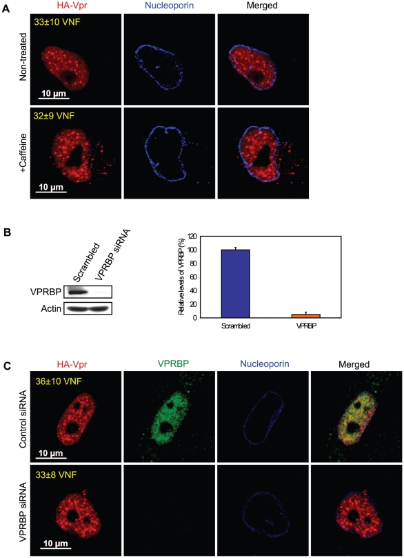
Figure 3. Formation of Vpr nuclear foci is independent of ATR activation and of the recruitment of VPRBP.
A) HeLa cells were pre-treated with 2.5 mM caffeine for 1 hour and then transduced with lentiviral vectors co-expressing GFP and HA-Vpr (WPI-HA-Vpr) or expressing GFP alone (WPI). One day after transduction, cells were fixed, permeabilized, and stained with antibodies against HA (red) and nucleoporin (blue). Images were acquired by confocal microscopy. Images shown are representative of multiple fields. Averages of the number of Vpr nuclear foci (VNF) per cell and corresponding standard deviations are shown. B) HeLa cells were transfected with control scrambled siRNA or siRNA targeting VPRBP. Forty-eight hours after transfection, cells were lysed and expression of VPRBP was monitored by western blot. VPRBP and actin were detected using rabbit polyclonal antibodies. Levels of VPRBP were monitored by computer-assisted densitometry and normalized for actin levels. The means (expressed as percentage relative to levels of VPRBP in scrambled siRNA-transfected cells (100%)) of three independent experiments are depicted in the graph on the right panel. C) HeLa cells were transfected with control scrambled siRNA or siRNA targeting VPRBP. Twenty-four hours after transfection, cells were transduced with lentiviral vectors co-expressing GFP and HA-Vpr (WPI-HA-Vpr) or expressing GFP alone (WPI). One day after transduction, cells were fixed, permeabilized, and stained with antibodies against HA (red), nucleoporin (blue) and VPRBP (green). Images were acquired by confocal microscopy. Images shown are representative of multiple fields. Averages of the number of Vpr nuclear foci (VNF) per cell and corresponding standard deviations are shown.

