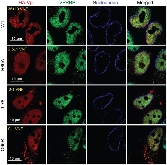
Figure 4. Analysis of the capacity of Vpr mutants to form nuclear foci.
HeLa cells were transfected with plasmids expressing HA-tagged Vpr (WT), Vpr (Q65R), Vpr (R80A), and Vpr (1–78). Forty-eight hours after transfection, cells were fixed, permeabilized, and stained with antibodies against HA (red), nucleoporin (blue) and VPRBP (green). Images were acquired by confocal microscopy. Images shown are representative of multiple fields. Averages of the number of Vpr nuclear foci (VNF) per cell and corresponding standard deviations are shown.

