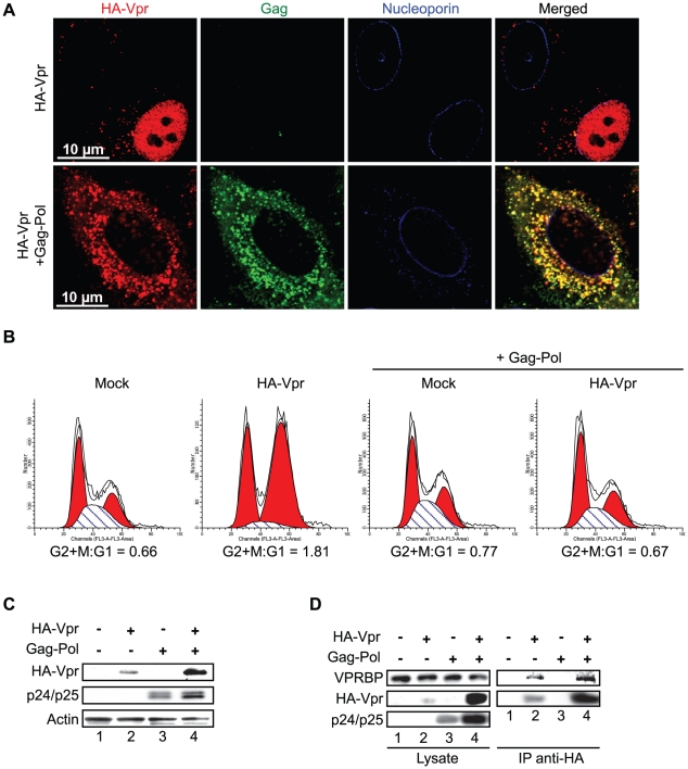
Figure 5. Cytoplasmic sequestration of Vpr abrogates foci formation and G2 arrest.
A) HeLa cells were co-transfected with the packaging plasmid psPAX2 encoding Gag-Pol, Tat, and Rev and with a HA-Vpr-expressing plasmid or appropriate empty plasmid control. Two days after transfection, cells were fixed, permeabilized, and stained with antibodies against HA (red), nucleoporin (blue) and p24 (green). Images were acquired by confocal microscopy. Images shown are representative of multiple fields. B) HEK293T cells were co-transfected with plasmids expressing GFP, HA-Vpr and Gag-Pol (psPAX2) or with an empty plasmid control as indicated. Forty-eight hours after transfection, cell cycle analysis was performed by flow cytometry using propidium iodide staining. Percentages of G1 and G2/M cell populations were determined using the ModFit software. C) Expression of HA-Vpr and p24 was monitored by western blot using specific monoclonal antibodies. Actin was detected using a rabbit polyclonal antibody. D) HEK293T cells were transfected as in B). Two days after transfection, cells were lysed and subjected to anti-HA immunoprecipitation as described in Materials and Methods. HA-Vpr, p24 or VPRBP levels were evaluated in cell lysates and immunocomplexes. HA-Vpr and p24 were detected using specific monoclonal antibodies. VPRBP was detected using a rabbit polyclonal antibody.

