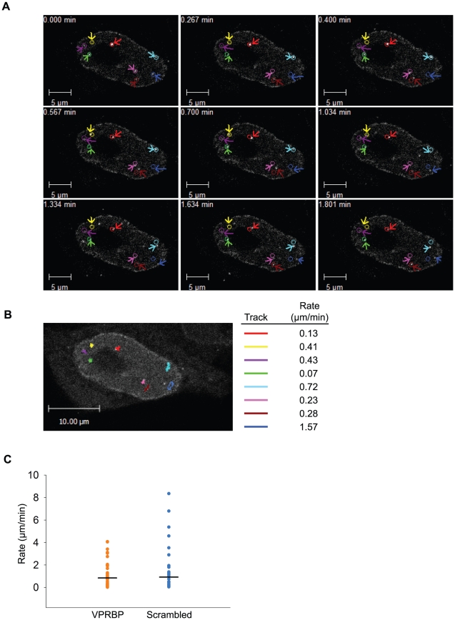
Figure 10. Vpr nuclear foci are mobile nuclear bodies.
A) HeLa cells were transfected with a plasmid expressing eYFP-Vpr WT. Two days after transfection, the location of eYFP-Vpr was monitored by time-lapse confocal microscopy in living cells. Images were acquired with a 63× objective at intervals of 2 seconds for two minutes. Representative images taken at time points spanning the period of acquisition are shown. The initial positions of Vpr foci are depicted by colored circles. The actual positions of foci are indicated with colored arrows. B) Vpr foci in images acquired in A) were tracked using the Volocity software v.5.2.1. Movement tracks of foci are depicted in colors on the picture. Rates of displacement for each focus are indicated at the right of the picture. Please note that some foci could not be tracked for the full time of acquisition because they were migrating out of the focal plane. C) HeLa cells were first transfected with scrambled siRNA or siRNA targeting VPRBP. Twenty-four hours after transfection, cells were transfected with a plasmid expressing eYFP-Vpr. The location of eYFP-Vpr foci was determined as in A) and rates of displacement were calculated as in B). Rates of displacement of individual foci are shown in a dot plot. Averages of displacement rates are indicated as horizontal bars.

