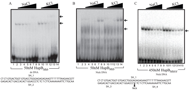
Figure 2. HupBMtb binds specifically to nick DNA under high salt conditions.
Comparative gel retardation analysis showing the binding of (A) 50 nM of HupBMtb under increasing salt concentration (0, 10, 50, 100, 150, 200 and 250 mM) of either NaCl (lanes 1–7) or KCl (lanes 8–14) as indicated in figure, to DNA (Table 2) ds or (B) nick DNA. (C) 450 nM of HupBMtbN binding to nick DNA under increasing salt concentration (0, 10, 50, 100, 150, 200 and 250 mM) of either NaCl or KCl as indicated in figure. Position of the nick in the DNA is marked by an arrowhead.

