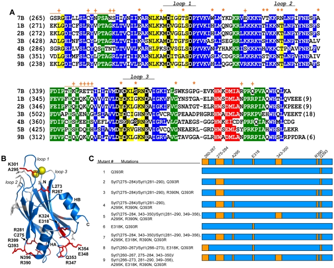
Figure 2. Sequence differences between the C2B domains of synaptotagmins-1 and -7.
(A) Sequence alignment of the C2B domains from selected rat synaptotagmin isoforms illustrating the similarities and differences among them. The first residue number of each sequence in each line is indicated in parenthesis. Conserved residues are color-coded: blue, β-strands; red, helix HA; yellow, top loops; green, bottom loops; black, Ca2+ ligands. The positions of the three Ca2+-binding loops (loops 1–3) are indicated. Residues that are identical in the C2B domains of synaptotagmins-1, -2 and -9 (the three isoforms that can support fast neurotransmitter release in cortical synapses [8]), but are different in the C2B domain of synaptotagmin-7, are indicated by a + sign if they were mutated in the Syt1/7 chimeras (see panel C), or by a * otherwise. The orange lines above the synaptotagmin-7 C2B domain sequence indicate the positions of fragments in the linker or loop sequences that were replaced with those of synaptotagmin-1 in the Syt1/7 chimeras (see panel C). (B) Ribbon diagram of the NMR structure of the synaptotagmin-1 C2B domain [17] illustrating the positions of selected residues mutated in the Syt1/7 chimers (shown as red stick models). The corresponding residue names (in single letter amino acid code) and numbers in synaptotagmin-1 are indicated above, and those in synaptotagmin-7 are indicated below. The bound Ca2+ ions are shown as yellow spheres. The positions of the Ca2+-binding loops and the two helices (HA and HB) are indicated. (C) Summary of chimeric Syt1/7 mutants used in this study. As illustrated in Figure 1C, Syt1/7 WT is composed of residues 1–265 of synaptotagmin-1 and the WT C2B domain (residues 260–403) of synaptotagmin-7. Each of the Syt1/7 mutants is composed of the same residues of synaptotagmin-1 (residues 1–265) and synaptotagmin-7 (residues 260–403) (represented in blue in the diagrams), but with the indicated mutations (illustrated by the orange boxes in the diagrams). The residue numbers of mutations are based on the respective WT synaptotagmins-1 and -7. In addition to point mutations, several stretches of residues in the synaptotagmin-7 C2B domain were changed to the corresponding residues in the synaptotagmin-1 C2B domain. These changes are listed as Syt7(residues)/Syt1(residues). For example, in Syt1/7 mutant #5, the synaptotagmin-7 residues 275–284 and 343–350 were replaced by the synaptotagmin-1 residues 281–290 and 349–356, respectively.

