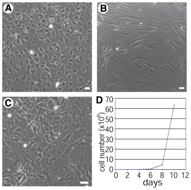
Figure 1.
JK1 cells exhibit distinct morphological differences from mouse testis stroma (MTS) and mouse embryonic fibroblasts (MEFs). (A–C): Phase-contrast images of a confluent monolayer of JK1 (A), MTS (B), and MEFs (C) showed that JK1 cells are adherent cells containing one or multiple nuclei, whereas MTS and MEFs contain more spindle-shaped cells (size bars = 50 μm). (D): JK1 cells were stained with trypan blue and counted using a hemocytometer over the course of 1 week, exhibiting exponential growth.

