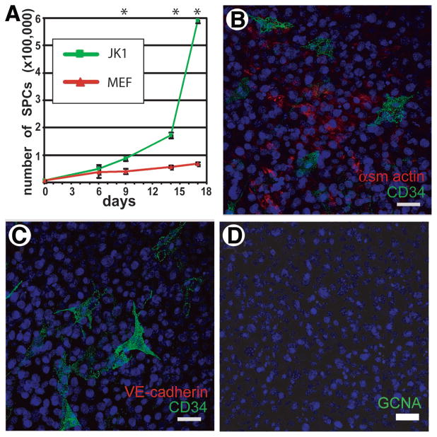
Figure 4.
JK1 cells enhance proliferation of spermatogonial progenitor cells (SPCs) and express markers associated with peritubular myoid cells. (A): The growth rates (mean ± SD) of SPCs on JK1 cells and MEFs were compared over the course of 2.5 weeks (n = 3; * indicates p < .05 by Student’s t test). (B–C): Coimmunostaining for CD34 (green) and either α-smooth muscle actin (red, B) or VE-cadherin (red, C) indicated that the JK1 cells contained cells positive for either αSMA or CD34 but not both, whereas VE-cadherin staining was completely negative. (D): JK1 cells also did not contain GCNA (red)-positive cells, indicating that they probably did not originate from GCNA-positive germ cells or VE-cadherin-positive endothelial cells in the testis. Nuclear counterstain is blue. All size bars = 50 μm. Abbreviations: MEF, mouse embryonic fibroblast.

