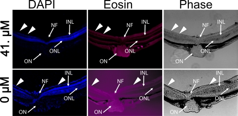
Figure 5.
Histopathologic examination of radial sections through the optic nerve head of mice eyes 7 weeks after intravitreal injection of balanced salt solution or 41 μM (100× MIC90) of C+balanced salt solution. Blue: DAPI-stained nuclei in various cell layers; red: eosin-stained cell membranes prominent in the plexiform layers. No retinal abnormalities were noted in the retinas injected with balanced salt solution alone or 0.41 to 4.1 μM of C+balanced salt solution. Eyes injected with 41 μM of C+balanced salt solution showed loss of cells in the ganglion cell layer (arrowheads). ONL, outer nuclear layer; INL, inner nuclear layer; GCL, ganglion cell layer, NF, nerve fiber; ON, optic nerve. Scale bar, 20 μm.

