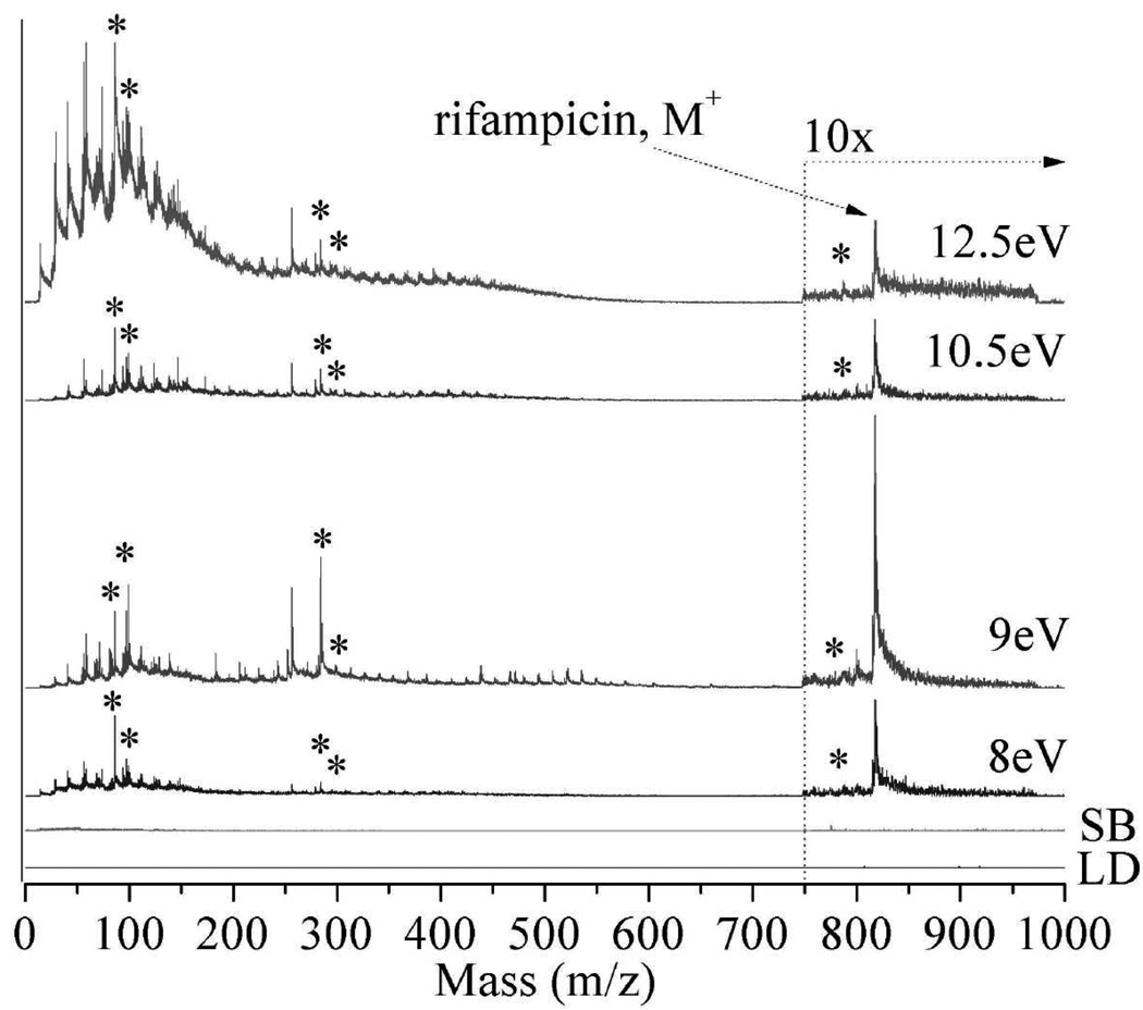
Figure 2.
Laser desorption postionization mass spectrometry (LDPI-MS) of neat rifampicin at photon energies of 8.0 – 12.5 eV (indicated on each spectrum). Also shown are (LD) direct ionization by laser desorption without vacuum ultraviolet (VUV) radiation and (SB) synchrotron background at 12.5 eV photon energy (no LD). Ion signal is shown after correction for photon flux variation with photon energy. Asterisks indicate known rifampicin fragments. Spectra here and below are artificially offset to avoid peak overlap.

