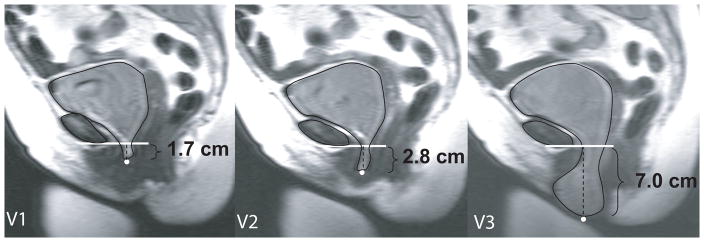
Figure 1. Increase in cystocele size with subsequent Valsalva efforts.
Demonstration that one (V1), or even two (V2), Valsalva attempts does not fully capture the extent of the prolapse. Data for this individual is seen in Figure 2 with white squares at reference points (red squares on color image).

