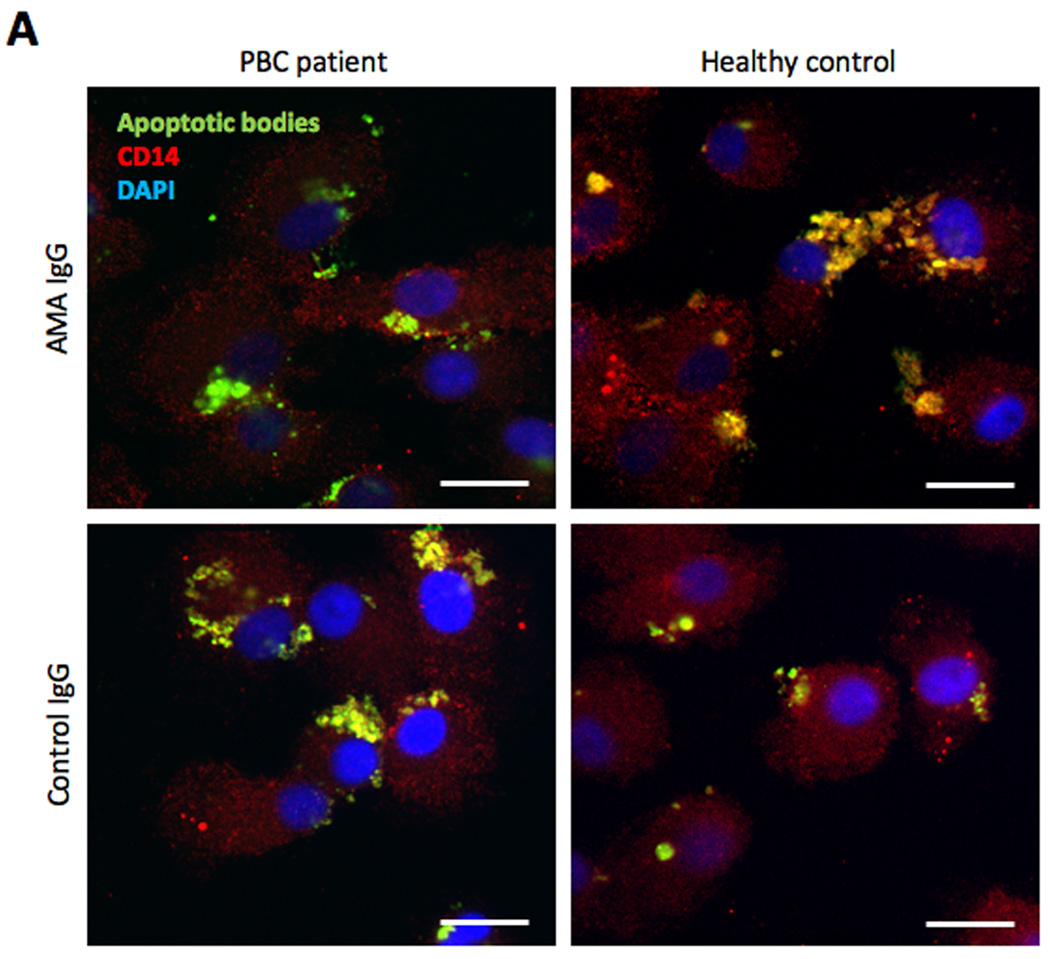
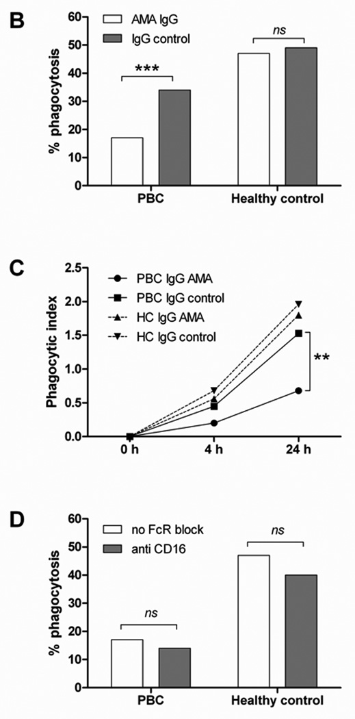
Figure 6. Uptake of apoptotic bodies.
(A) Confocal imaging of MDMΦ from a representative patient with PBC and an unaffected control co-incubated for 24 h with CFSE-labeled apoptotic bodies from HIBEC in the presence of human AMA-IgG or control- IgG. After culture the cells were fixed and stained with PE-conjugated anti-human CD14 (red), DAPI was used to stain the nucleus (blue), CFSE stained apoptotic bodies are shown in green. In all four conditions, apoptotic bodies (green) are located inside the cells, which indicate that MDMΦ actively engulfed apoptotic bodies after 24 h (arrows). Scale bar 20 µm. (B) Comparison of phagocytic efficacy, expressed as percentage of phagocytosis, of MDMΦ from PBC and controls; MDMΦ from PBC had a reduced uptake of HIBEC apoptotic bodies in the presence of AMA (17% vs 47%, ***p<0.001), this difference was not noted when IgG control was used (34% vs 49%, p = ns). Percentage of phagocytosis was calculated by counting the number of macrophages that had ingested at least one HIBEC apoptotic body. (Fisher's Exact Test). (C) The phagocytic index (PI) was expressed as percentage of phagocytosis multiplied by the mean number of phagocytosed bodies per macrophage (ABMΦ): PI = (% phagocytosis x mean ABMΦ/100) and was evaluated at 0, 4 and 24 h of incubation. ** p< 0.01 (D) The ability of MDMΦ from PBC and controls to uptake HIBEC apoptotic bodies, expressed as percentage of phagocytosis, was studied in presence of AMA, with or without FcγR block. No significant difference (ns) in the percentage of phagocytosis was observed when FcγR was blocked in both patients and controls. Statistical difference was determined by Fisher's Exact Test.


