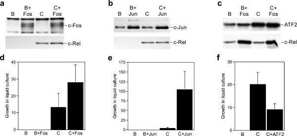
Figure 4.
Differential effect of enhanced AP-1 expression on the transformation potential of c-Rel. (a–c) Whole cell lysates were prepared from CEFs expressing control BIS retroviruses (B), BIS viruses expressing c-Rel (C), c-Fos, c-Jun, and ATF2 alone, or viruses that co-expressed c-Rel with each of the AP-1 family members. The expression of c-Fos, c-Jun, ATF2, and c-Rel was analyzed by Western blot. Proteins detected by the antisera are indicated on the right of each panel. (d–f) The viruses described above were used to infect splenic lymphocytes from three-week old chickens. Three days later, infected cells were serially diluted in 96-well plates and growth was scored microscopically after ten days. The average and standard deviation of three independent experiments is shown.

