
Figure 6.
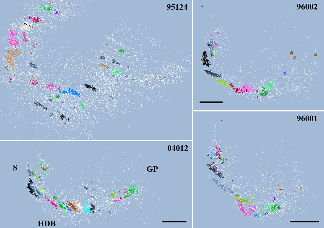
Distribution of cholinergic cells (unassigned in white) and clusters (in various colors) viewed from approximately the same angle in four different rats. Case 95124 were cut in horizontal plane; the three other brains were cut coronal. Cluster parameters are summarized in Table 1. The location of the septum (S), horizontal limb of the diagonal band (HDB) and globus pallidus (GP) are indicated. For better view of the clusters, section outlines are omitted. Scale in case #04012: 0.85 mm; case 96002: 1mm; case 96001: 1.5 mm.