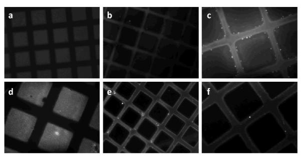
Figure 8.

Fluorescence microscopy images of a photo-patterned dextranized surface on polyurethane (PU): (a) before and (b) and (c) after adsorption of human albumin (HA) in PBS for 1h and on PS film: (d) before and (e) and (f) after adsorption of human albumin (HA) in PBS for 1h. Prior to adsorption of HA, dextranized regions (squares) show fluorescence (white regions) due to the aryl group (AZBC) used to photoactivate the dextran. Upon HA adsorption, bright fluorescence appears on the non-dextranized regions, due to localized adsorption of fluorescently labeled HA on PU and PS regions. The pattern dimensions are 90×90 μm2 separated by 35 μm. The image magnifications are 20x for (a), (b) and (e) and 40x for (c), (d) and (f).
