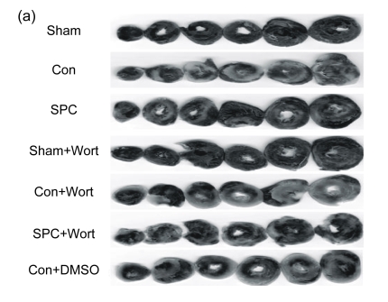
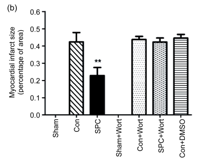
Fig. 3.
Myocardial infarct size in each experimental group
(a) Representative photographs of heart slices with 1% (w/v) triphenyltetrazolium chloride staining in each group; (b) Average infarct size in six hearts for each group. The size of the infarcted myocardium was calculated as an area ratio (necrotic area/total area of the myocardium×100%). ** P<0.01 compared with the Con group


