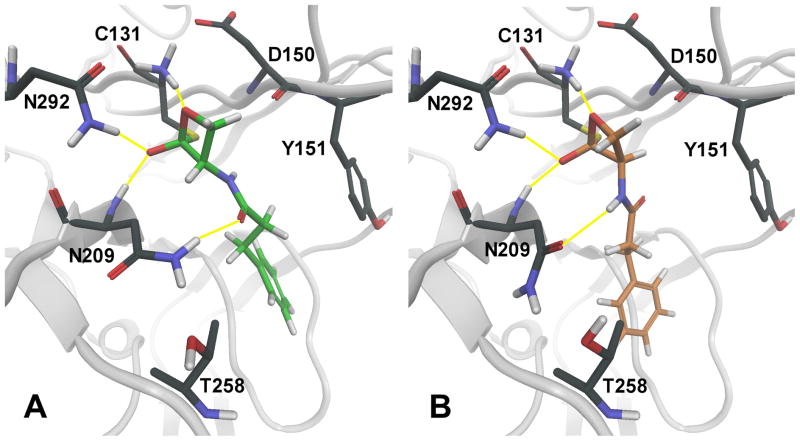
Figure 2.
Representation of the putative tetrahedral intermediates resulting from the nucleophilic attack of catalytic cysteine 131 onto the lactone carbonyl of 7a (A, green carbons) or 8a (B, orange carbons). The backbone of the NAAA model, built by comparative modeling, is represented in grey. Hydrogen bonds between enzyme residues and the inhibitor are symbolized by yellow lines. Standard-atom color codes: black: carbon; red: oxygen; blue: nitrogen; white: hydrogen; yellow: sulfur.

