
Figure 1.
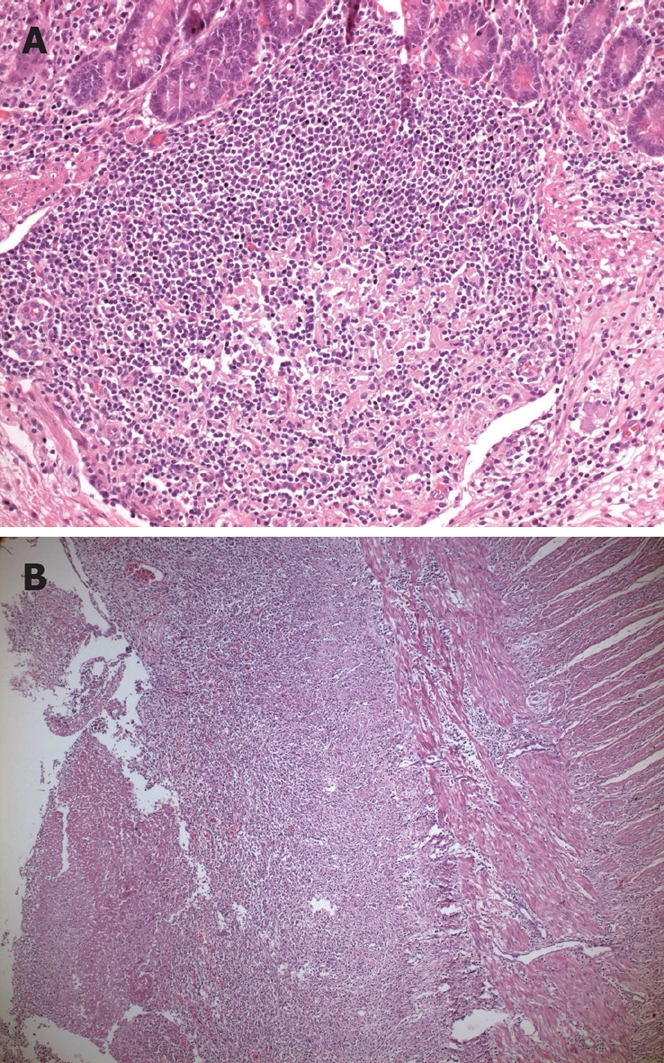
Histopathologic view of typhoid lesions. A: Typhoid nodule, there are macrophages containing bacteria, red blood cells, and nuclear debris from small nodular aggregates in Peyer’s patches (HE stain, × 20 objective); B: Typhoid ulceration (HE stain, × 5 objective).