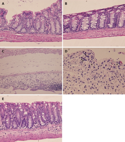
Figure 3.
Histological analysis of mice. A: Colon section from control (water treated) animals showing normal colon tissue architecture (HE, × 400); B: Colon section from Arctium lappa L. (AL)-treated animals showing normal histological architecture with no inflammatory cell infiltration, edema or crypt abscesses (HE, × 400); C, D: Colon section from dextran sulfate sodium (DSS)-treated animals showing severe submucosal erosion with edema, ulceration, inflammatory cell infiltration (indicated with arrows) and crypt abscesses as well as epithelioglandular hyperplasia (C: HE, × 200, D: HE, × 400); E: Colon section from animals challenged with DSS after prior treatment with AL showing normal histological architecture with slight inflammatory cell infiltration and no submucosal edema or abnormality of crypt cells (HE, × 400). All the results are representative of 3 independent experiments.

