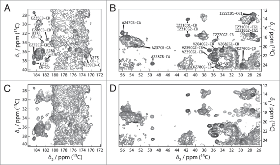
Figure 4.
(A and B) Extracts of the 100 ms DARR spectra of HET-s(218-289) (blue) and HET-s(black). All peaks of the prion domain are present in the spectrum of the HET-s fibrils. (C and D) Extracts of the 100 ms DARR spectra of HET-s(1-227) (red) and HET-s(black). The structure of the prion domain is the same as in HET-s(218-289) while the globular domain looses its well-defined teritiary structure.7

