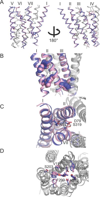
Figure 5.
Modeling the structural divergence between rhodopsin and β2AR for helices II, III, and V. The β2AR model (magenta) generated by the restraint sets β2AR/pred/struct (see Table I for annotation) is superimposed to the rhodopsin (green) and the β2AR (blue) crystal structures using backbone atoms of helices II, III, and V.

