
Fig. 9.
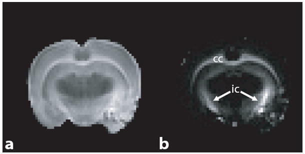
Normalized images from chromated brain at TE = 6.5 ms (a) and 71.5 ms (b). In (b), the signal from both myelin and extra-axonal water has nearly decayed to baseline, while residual signal from intra-axonal water remains. Note, that substantial signal remains within the corpus callosum (cc) and internal capsule (ic).