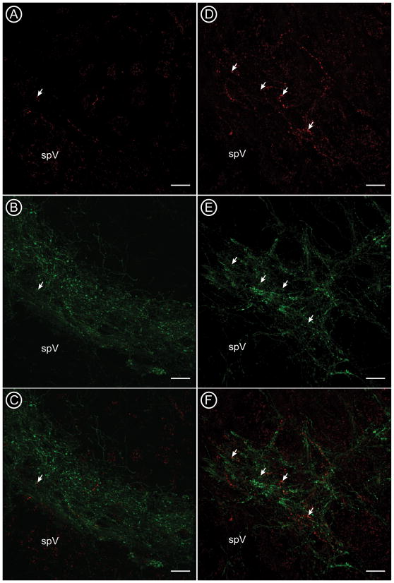
Figure 2.
Confocal micrographs show the distribution of corneal afferents labeled with CTb (A, D; red) and nociceptive CGRP-containing afferents (B, E; green) at the caudal (A-C) and rostral (D-F) boundaries of ventrolateral Vc. Some CTb-labeled varicosities also contained CGRP (arrows). spV = spinal trigeminal tract. Scale bars = 20 μm.

