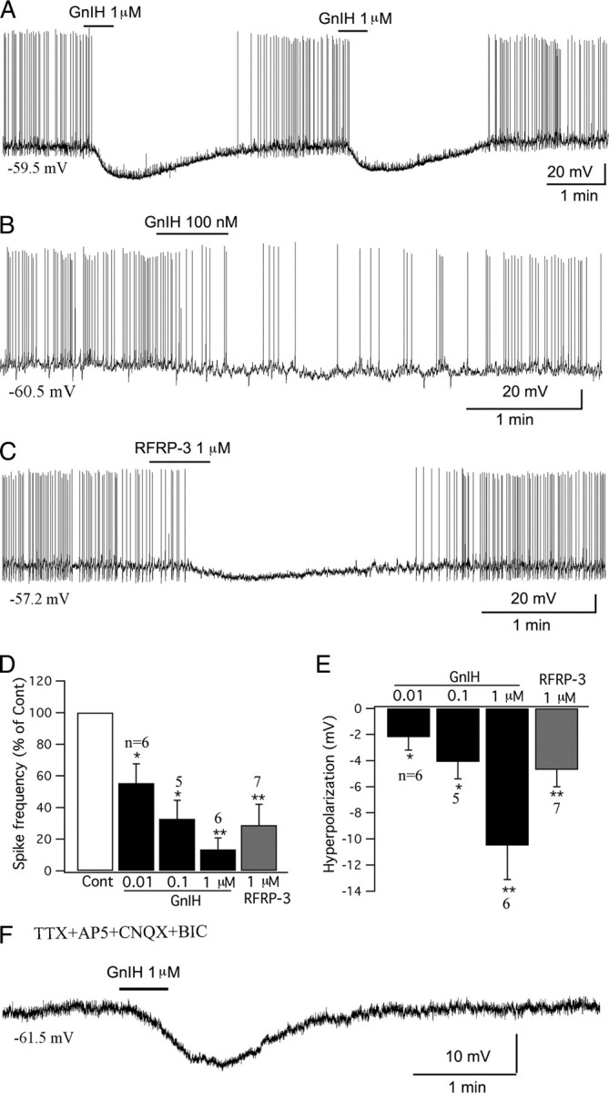
Figure 8.

GnIH inhibits POMC cells in arcuate nucleus. A, A typical trace showing that GnIH (1 μm) inhibited the firing and hyperpolarized the membrane potential of a POMC cell. The effect was repeatable with four successive applications of GnIH; only the first two applications are shown here. B, GnIH (100 nm) inhibited a POMC cell. C, The mammalian analog of GnIH, RFRP-3 (1 μm) depressed a POMC cell. D, Bar graph showing the dose-dependent effects of GnIH (0.01–1 μm) and RFRP-3 (1 μm) on the spike frequency of POMC cells (control, 100%; *p < 0.05, **p < 0.01 vs Cont; ANOVA). E, Bar graph showing the dose-dependent hyperpolarization that GnIH (0.01–1 μm) and RFRP-3 (1 μm) had on POMC cells (*p < 0.05, **p < 0.01 vs Cont; ANOVA). Error bars indicate SEM. F, Trace showing that GnIH (1 μm) hyperpolarized a POMC cell in the presence of TTX (0.5 μm), AP5 (50 μm), CNQX (10 μm), and BIC (30 μm) in bath solution, indicating a direct effect.
